Translate this page into:
Iatrogenic Pseudoaneurysm of Intervalvular Fibrosa – A Case Report
*Corresponding author: Rajiv K. Gupta, Department of Cardiothoracic Surgery, Dayanand Medical College, Ludhiana, Punjab, India. drrajivkr@yahoo.co.in
-
Received: ,
Accepted: ,
How to cite this article: Gupta RK, Dhindsa D, Sindwani R, Kapoor S. Iatrogenic Pseudoaneurysm of Intervalvular Fibrosa – A Case Report. Indian J Cardiovasc Dis Women. 2024;9:188-90. doi: 10.25259/IJCDW_49_2023
Abstract
Pseudoaneurysm of aorto-mitral intervalvular fibrosa (P-AMIVF) is a rare fatal complication of endocarditis; however, balloon aortic valvuloplasty has been associated with this condition. In the absence of complications, this entity is usually asymptomatic. Trans-thoracic and trans-esophageal echocardiography are the mainstay to assess the severity of valve lesion and myocardial abscess in this hidden region. The 38-year-old patient diagnosed with critical calcified bicuspid aortic valve stenosis; dilatation of aortic root and aneurysmal aorto-mitral curtain with moderate mitral regurgitation was treated successfully. We share our views and challenges related to mural endocarditis, myocardial abscesses, and P-AMIVF.
Keywords
Infective endocarditis
Pseudoaneurysm
Aorto-mitral intervalvular fibrosa
Transthoracic
Transesophageal echocardiography
Anterior mitral leaflet
Posterior mitral leaflet
INTRODUCTION
Aorto-mitral intervalvular fibrosa (AMIVF) is a hidden avascular membrane between non-coronary cusp of aortic valve and anterior mitral leaflet (AML) of mitral valve.[1] It is essential for maintaining functional and anatomic integrity. Pseudoaneurysm of the aortomitral intervalvular fibrosa (P-AMIVF) may be primary or myocardial abscesses secondary to infected mural thrombus, intracardiac devices, prosthetic, and valvular infective endocarditis.[2,3]
Systemic embolization, abscess and fistula formation, papillary muscle or chordae damage, and cardiac perforation are the potential complication of mural endocarditis.[4]
CASE REPORT
A 38-year-old patient admitted with repeated syncopal attacks and decreased vision in left eye. Two years ago, the patient underwent balloon aortic valvuloplasty (BAV) and subsequently developed fever and acute abdominal pain. The patient was treated with intravenous antibiotics for 8 weeks. Transthoracic echocardiography (TTE) in the parasternal long and short axis, revealed calcified bicuspid stenotic aortic valve with subannular aortic dilatation in the region of intervalvular fibrosa involving the AML.
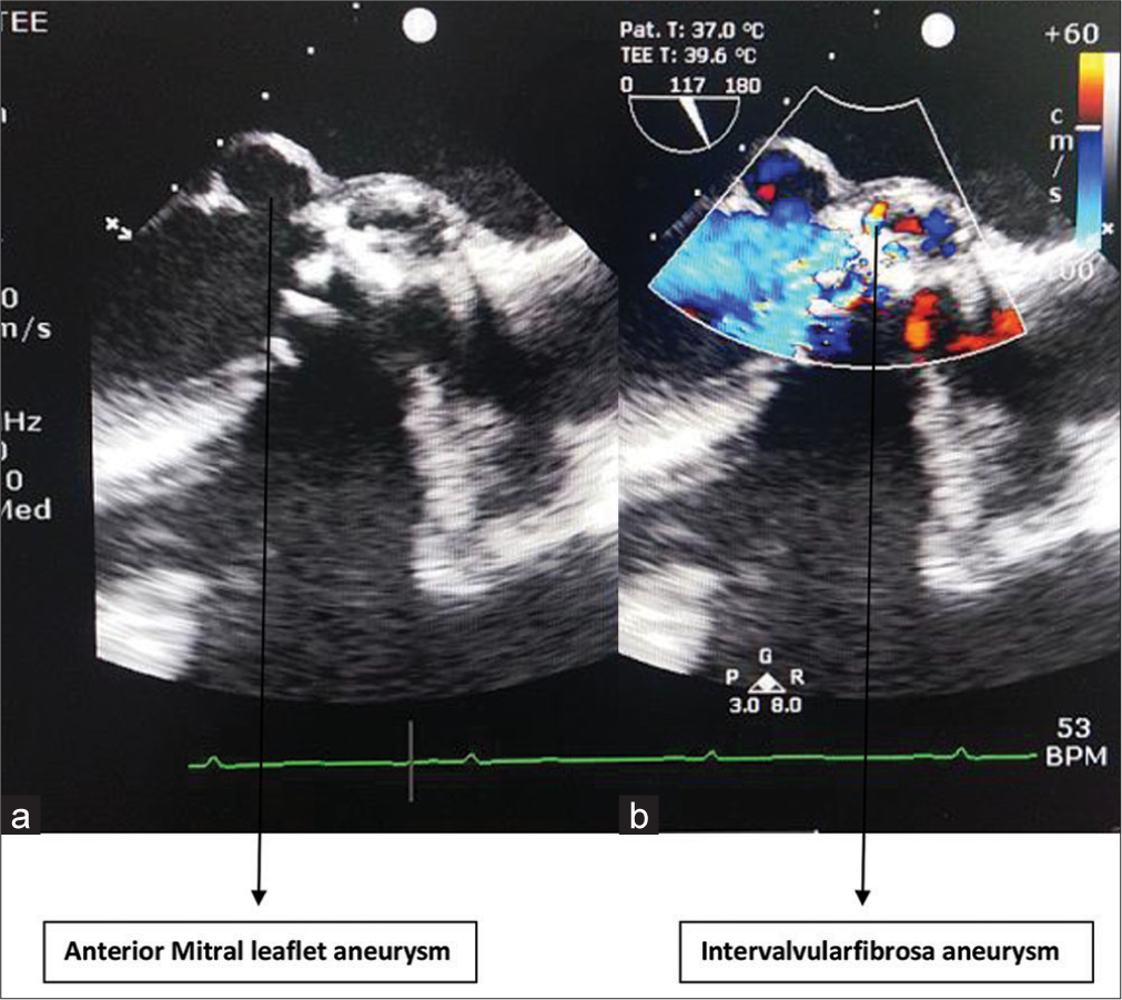
Trans-esophageal echocardiography (TEE) revealed in the mid-esophageal three-chamber view echo free space at posterior aortic root and aneurysm in the aorto-mitral curtain [Figure 1].

- (a) Anterior mitral leaflet aneurysm and (b) intervalvular fibrosa aneurysm.
Operative findings were of calcified bicuspid aortic valve; and after excesing the aortic leaflets; aneurysmal opening in the subannular posterior aortic root in the area of intervalvular fibrosa was visible. There was a punched out area in the middle of the AML body.
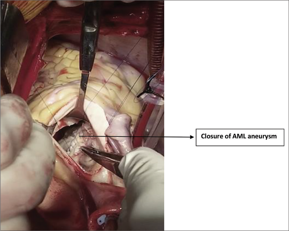
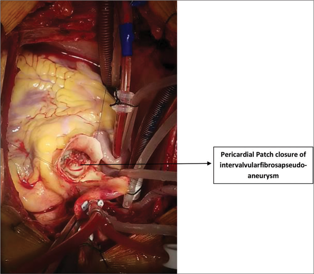
Mitral valve was approached through a transatrial approach. A aneurysmal sac was visible in the middle of fibrous trigone (A2 area). The sac was excised and sutured directly and posterior mitral leaflet (PML) stabilized with annuloplasty band. The punched out area on AML was closed with pericardial patch using proline5-o suture [Figures 2 and 3]. Aortic valve was replaced with 21 mm St. Jude Valve. Patient was weaned off cardiopulmonary bypass successfully and the post-operative period was uneventful.

- Closure of anterior mitral leaflet (AML) aneurysm.

- Pericardial patch closure of intervalvular fibrosa pseudoaneurysm.
DISCUSSION
This rare anomaly presented to us following history of BAV followed by infective endocarditis leading to P-AMIVF. The aortomitral curtain is avascular area, juxtaposition with epicardial wedge of fat lying between aorta and left atrium posteriorly.[5]
In 1924, mural endocarditis involving non-valvular endocardium was described first and is similar to valvular endocarditis clinically. The two can be differentiated with imaging techniques.[6]
The various causes of non-valvular infective endocarditis are secondary to infected mural thrombus, intracardiac devices, cardiac tumors, and congenital defects.[5]
In this case following BAV 2 years ago, the patient developed high grade fever with abdominal pain. Treatment comprised of antibiotics for 2 months and leading to relief of symptoms. The high velocity regurgitant jet caused endothelial disruption over the AML creating a punched out area over the AML. This endothelial disruption attracts platelet and fibrous deposition thus creating a nidus for bacterial infection. This leads on to mural infective endocarditis of the AMIVF forming P-AMIVF. The timely intervention with intravenous antibiotics prevented the abscess formation, thus contained rupture.
The main imaging studies are TTE and TEE. Doppler color flow mapping reveals pulsatile echo free-pouch with both systolic expansion and diastolic collapse. TEE is superior to TTE in the evaluation of P-AMIVF which is located posterior to cardiac chambers. TEE can detect and localize the subaortic lesion such as abscess; intervalvular pseudoaneurysm; and its communication and perforation of AML.[7]
CONCLUSION
Early surgical intervention is a well established treatment of valvular endocarditis with myocardial abscess and P-AMIVF. In this case, the P-AMIVF was missed for the first time thus delayed presentation with a thromboembolic event in the form of decreased vision of the left eye. The only option available for myocardial abscess and for P-AMIVF is surgery.
Ethical approval
The Institutional Review Board approval is not required.
Declaration of patient consent
The authors certify that they have obtained all appropriate patient consent.
Conflicts of interest
There are no conflicts of interest.
Use of artificial intelligence (AI)-assisted technology for manuscript preparation
The authors confirm that there was no use of artificial intelligence (AI)-assisted technology for assisting in the writing or editing of the manuscript and no images were manipulated using AI.
Financial support and sponsorship
Nil.
References
- Pseudoaneurysm of the Mitral-aortic Intervalvular Fibrosa (MAIVF): A Comprehensive Review. J Am Soc Echocardiogr. 2010;23:1009-18.
- [CrossRef] [PubMed] [Google Scholar]
- Challenges in Infective Endocarditis. J Am Coll Cardiol. 2017;69:325-44.
- [CrossRef] [PubMed] [Google Scholar]
- Fibrous Skeleton of the Heart: Anatomic Overview and Evaluation of Pathologic Conditions with CT and MR Imagining. Ragiographics. 2017;37:1330-51.
- [CrossRef] [PubMed] [Google Scholar]
- Mullisite Infective Endocarditis with Mural Vegetation in the Right Atrium and Right Ventricle. Circulation. 2011;123:457-8.
- [CrossRef] [PubMed] [Google Scholar]
- Mitral-Aortic Intervalvular Fibrosa. J Am Coll Cardiol Case Rep. 2020;2:1217-9.
- [CrossRef] [PubMed] [Google Scholar]
- Bacterial Mural Endocarditis. A Case Series. Heart Lung Circ. 2014;23:e172-9.
- [CrossRef] [PubMed] [Google Scholar]
- Pseudoaneurysm of Mitral-aortic Intervalvular Fibrosa. Int J Cardiol. 2013;166:2-7.
- [CrossRef] [PubMed] [Google Scholar]


