Generic selectors
Exact matches only
Search in title
Search in content
Post Type Selectors
Search in posts
Search in pages
Filter by Categories
Abstracts
Cardiovascular, Case Report
Cardiovascular, Commentary
Cardiovascular, Editorial
Cardiovascular, Guest Editorial
Cardiovascular, Images in Cardiology
Cardiovascular, Interventional Round
Cardiovascular, Original Article
Cardiovascular, Perspective Review
Cardiovascular, Preface
Cardiovascular, Review Article
Cardiovascular, Student’s Corner
Case Report
Case Report, Cardiovascular
Case Reports
Case Series, Cardiovascular
Clinical Discussion
Clinical Rounds
CPC
Current Issue
Debate
Dedication
Editorial
Editorial Cardiovascular
Editorial, Commentary
Editorial, From the Publisher’s Desk
Expert Comments
Expert's Opinion
Genetic Autopsy
Genetics Autopsy
Guest Editorial, Cardiovascular
Image in Cardiology
Images in Cardiology
Images in Cardiology, Cardiovascular
Interventional Round
Interventional Round, Cardiovascular
Interventional Rounds
Letter to Editor
Letter to the Editor
Media and news
Original Article
Original Article, Cardiovascular
Original Article, Cardiovascular Health
Practice in Medicine
Preface
Review Article
Review Article, Cardiovascular
Scientific Paper
Short Communication
Student's Corner
Supplementary
Supplemetary
WINCARS Activities
View/Download PDF

Translate this page into:

Case Report
02 (
03
); 054-055
doi:
10.1055/s-0037-1606851

2:1 Atrioventricular Conduction due to Congenital Long QT Syndrome

Department of Cardiology, Kasturba Hospitals, Mangalore, Karnataka, India
Department of Electrophysiology, Aayush Hospitals, Vijayawada, Andhra Pradesh, India

Soma Sekhar Ghanta, MD, DM, FEp Department of Electrophysiology, Aayush Hospitals Vijayawada 520008, Andhra Pradesh India somasekharghanta1978@gmail.com

Licence
This is an open access article published by Thieme under the terms of the Creative Commons Attribution-NonDerivative-NonCommercial License, permitting copying and reproduction so long as the original work is given appropriate credit. Contents may not be used for commercial purposes, or adapted, remixed, transformed or built upon. (https://creativecommons.org/licenses/by-nc-nd/4.0/)
Disclaimer:
This article was originally published by Women in Cardiology and Related Sciences and was migrated to Scientific Scholar after the change of Publisher.

Abstract

Abstract

Long QT syndrome patients can present as second-degree AV block due to prolongation of refractory period. We report a case of pseudo 2:1 AV block due to long QT syndrome in which treatment differs from conventional second-degree AV block because of uncommon presentation and poor prognosis with no consensus.

Keywords

long QT
second-degree AV block
2:1 AV conduction

Introduction

Congenital long QT syndrome is a rare disorder of prolongation of ventricular refractory period with prevalence of 1 in 5,000. It is a channelopathy with autosomal dominant and recessive inheritance with potassium, sodium, and calcium channel abnormalities. Patients with this disorder can present with syncope, ventricular tachyarrhythmias, and sudden cardiac arrest.

Electrocardiographic (ECG) manifestations of long QT syndrome are prolonged QT more than 470 milliseconds, T wave alternans, polymorphic ventricular tachycardia (VT), and, rarely, 2:1 atrioventricular (AV) conduction.1 2 3

Case Report

A 3-month-old child was referred to us with a history of shortness of breath for cardiac evaluation. She was evaluated with ECG revealing prolonged QT with QTc of 440 milliseconds and 2:1 AV conduction (Fig. 1A). Echocardiogram was normal with no evidence of structural abnormalities. Chest radiograph was also nondistinct. With suspicion of long QT, evaluation of sibling of 1 year 6 months was done, which revealed prolonged QT interval with QTc of 480 milliseconds (Fig. 1B), suggesting familial long QT syndrome. There was no family history of sudden cardiac arrest or syncope in siblings. She was started on propranolol for treatment of long QT and followed up.
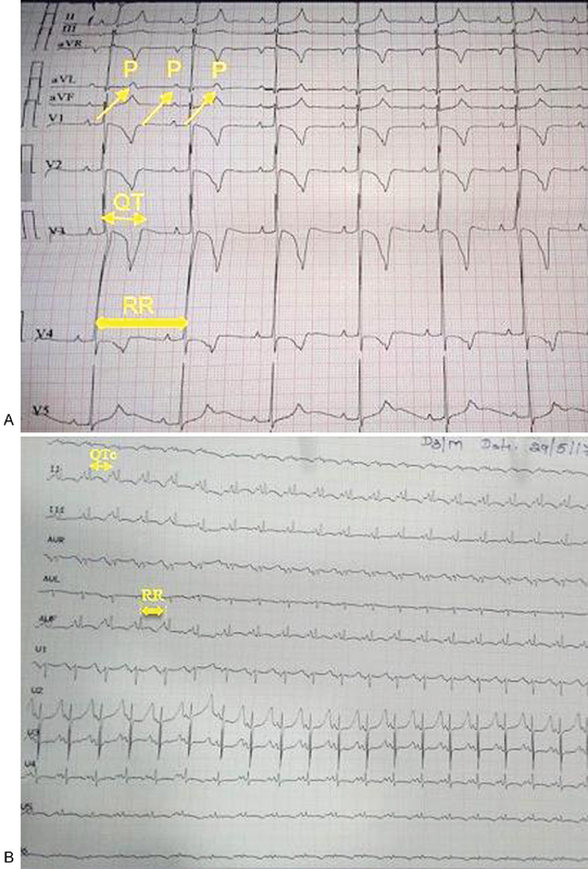
Fig. 1 (A) ECG of index patient showing arrows pointing P waves with alternate P on T waves and QT of 440 milliseconds and corrected QTc of 440 milliseconds. (B) ECG of sibling showing sinus tachycardia with RR interval of 440 milliseconds, QT interval of 320 milliseconds with QTc of 480 milliseconds.

Fig. 1 (A) ECG of index patient showing arrows pointing P waves with alternate P on T waves and QT of 440 milliseconds and corrected QTc of 440 milliseconds. (B) ECG of sibling showing sinus tachycardia with RR interval of 440 milliseconds, QT interval of 320 milliseconds with QTc of 480 milliseconds.

Discussion

Long QT syndrome is an inherited disorder of cardiac ion channel associated with prolongation of ventricular refractory period. It has predisposition to life-threatening ventricular arrhythmia causing sudden death. It can present as palpitations, presyncope, and aborted sudden cardiac arrest due to life-threatening ventricular arrhythmia predominantly polymorphic VT. When ventricular refractory period is extremely prolonged more than sinus rates, it may present as 2:1 AV conduction. When refractory period of ventricular myocardium is prolonged with preserved rate-dependent abbreviation of refractory period of Purkinje's network, sinus tachycardia manifests as 2:1 AV conduction.2 Though prolonged QTc is taken as 470 milliseconds as per Schwartz study, there are studies reported with mean QTc less than 440 milliseconds in Indian newborn infants.4 In our case, corrected QTc is 440 milliseconds and PP interval is also 440 milliseconds, which is relatively prolonged causing 2:1 AV conduction. Here β-blockers are mainstay of therapy in long QT, unlike other causes of 2:1 AV conduction. Genomic analysis is important in evaluating pediatric patients with suspicion of long QT syndrome for genetic counseling and guiding therapy as there is type-specific intervention, with long QT types 1 and 2 responding better to β blockers and type 3 responding better to mexiletine and, rarely, requiring pacing.1

References

  1. , , , . Second degree heart block associated with QT prolongation. Indian Pacing Electrophysiol J. 2010;10(02):96-98.
    [Google Scholar]
  2. , , . Pseudo 2:1 atrioventricular block and T wave alternans in the long QT syndromes. J Am Coll Cardiol. 1991;18(05):1363-1366.
    [Google Scholar]
  3. , , , , . A rare cause of 2:1 AV block: long QT syndrome. J Cardiovasc Electrophysiol. 2008;19(09):990.
    [Google Scholar]
  4. , , , et al. . QT interval in newborns of different ethnic origin: usefulness of neonatal ECG screening. Rev Esp Cardiol. 2008;61(09):980-982.
    [Google Scholar]

Fulltext Views
763

PDF downloads
517
View/Download PDF
Download Citations
BibTeX
RIS
Show Sections